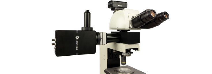
Focusing Modules for Mightex LED Collimator Sources
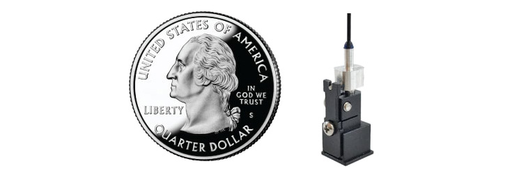
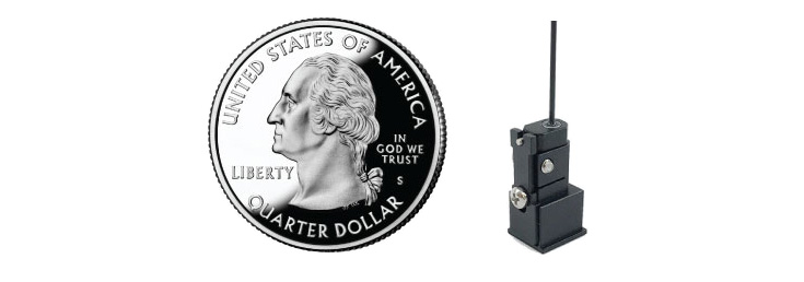
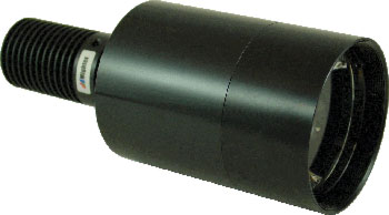
Focusing Modules for High-Power LED Collimator Sources:
- (P/N: ACC-LCS-F-11): Focusing Module for 11mm-clear-aperture LED Collimator Sources, Working Distance ~5mm;
- (P/N: ACC-LCS-F-22): Focusing Module for 22mm-clear-aperture LED Collimator Sources, Working Distance ~10mm;
- (P/N: ACC-LCS-F-22-50): Focusing Module for 22mm-clear-aperture LED Collimator Sources, Working Distance ~50mm;
- (P/N: ACC-LCS-F-22-100): Focusing Module for 22mm-clear-aperture LED Collimator Sources, Working Distance ~100mm;
- (P/N: ACC-LCS-F-38): Focusing Module for 38mm-clear-aperture LED Collimator Sources, Working Distance ~20mm;
- (P/N: ACC-LCS-F-48): Focusing Module for 48mm-clear-aperture LED Collimator Sources, Working Distance ~30mm.
IMPORTANT : (1) LED’s can ONLY be driven by a constant-current source, and NOT a voltage source (e.g. a battery, or a AC/DC power supply etc.); (2) Please always verify LED’s current rating first before applying current to the LED, and please always make sure NOT to apply current that is above the LED’s current rating.
About Mightex High Power LED Light Sources
Selecting a Microscopy LED Light Source
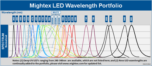
1. Select LED Wavelength
2. Select LED Configuration
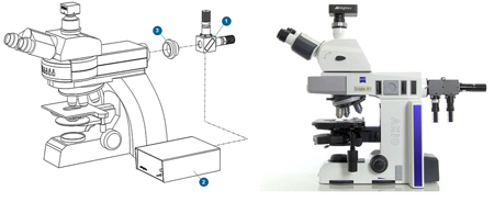
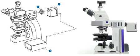
3. Select LED Controller
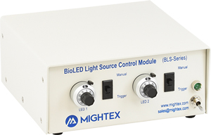
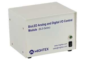
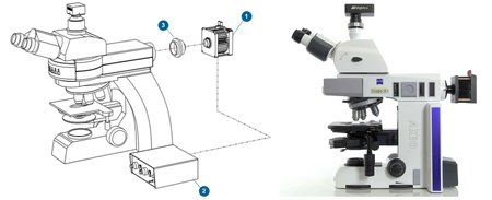
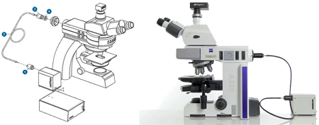
4. Microscope Coupling
Microscope adapters are available for all major manufacturers (Nikon, Olympus, Leica, Zeiss). If you have a different microscope, please contact us.

LED Application Examples
Abdelfattah AS et al., Bright and Photostable Chemigenetic Indicators for Extended In Vivo Voltage Imaging – Science (2019)
Abdelfattah et al., developed novel chemigenetic indicators for in vivo voltage imaging in neurons. To illuminate their indicators, the group used Mightex’s Microscope LEDs.
Crandall SR, et al., Infrabarrels Are Layer 6 Circuit Modules in the Barrel Cortex that Link Long-Range Inputs and Outputs – Cell Reports (2017)
In this work, Crandall et al., used Mightex’s Microscope LEDs to optically stimulating different neuronal pathways connecting to pyramidal neurons found in L6 of the whisker somatosensory cortex of mice.
Higgs MH, Measurement of Phase Resetting Curves Using Optogenetic Barrage Stimuli – Journal of Neuroscience Methods (2017)
To study phase resetting curves in vivo, Higgs and Wilson used Mightex’s Microscope LEDs to establish whether their novel method of optogenetic barrage stimulation would work during extracellular spike recordings (a recording method often used in in vivo experiments).
Also See:
Related Products:


