
")
-
Startseite

-
Produkte
-
Datenerfassung und -Analyse
-
Multielektroden-Aufnahmesysteme
-
Multikanal-Elektrodenträger für in-vivo-Ableitungen
- Floating Microelectrode Arrays (FMA)
Floating Microelectrode Arrays (FMA)


A sophisticated neural interface that is typically best suited for chronic recording and/or stimulation in large animal cortex, the FMA has also seen successful use in a variety of small animal and peripheral nerve studies. The low profile, small size, and great flexibility of the FMA make it ideal for the implantation of a large number of arrays simultaneously in the same animal, allowing a multitude of cortical locations to be targeted at the same time. Additionally, the length and impedance of individual electrodes within the same array are completely customizable, giving researchers the capability of recording multiple cortical layers and deep structures, or collecting LFP and single unit activity from the same cortical region. The FMA is also capable of remarkably long periods of chronic use, with over seven years of continual single unit recording having been reported from multiple arrays in non-human primate.
Characteristics
- Up to 36 channels per array.
- Custom impedance in each microelectrode.
- Custom length of each microelectrode of the array.
- Custom pedestals.
- Microelectrode impedance is individually customizable from anywhere between 5 kΩ to 5 MΩ.
- Small, low profile arrays allow many to be implanted into one animal (over 20 arrays in primate).
- Available in Platinum/Iridium and Pure Iridium, with activation of Iridium electrodes on request.
- Cable length up to 20 cm.
Design and Specifications
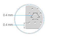
The Floating Microelectrode Array (FMA) is based on the concept of a lightweight platform populated with rigid microelectrodes and tethered to a nano connector by a thin, flexible and light-weight cable. The platform which houses the individual electrodes is fabricated from 125 µm thick alumina ceramic, and has 18 or 36 evenly distributed holes with 250 or 400 µm separations.
FMAs can be fabricated with varying electrode depths not typically achieved using planar silicon technology. Current fabrication techniques allow for individual electrode depths between 0.5 and 10 mm. Impedance values for individual electrodes within the array may be specified from 5.0 kΩ to 5 MΩ.
The cable is fabricated from Parylene-C insulated 25 µm diameter gold wires, which are wound in a helix for maximum flexibility and strength. The entire cable is insulated with a thin coat non-restrictive silicone elastomer, ensuring biocompatibility, flexibility and strength.
Chronic FMA Assemblies
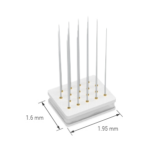
The array shown above is tethered by eighteen fine wires, 25 µm (0.001") in diameter, about ⅓ the size of a human hair. The gold wires are micro-welded to the shaft of the microelectrodes at the specified distance from the tip in order to establish the proper length of the electrode. The wires are sonically bonded to an Omnetics connector, which is used by leading headstage amplifier suppliers including Plexon, Ripple, TBSI, and Alpha Omega. These connectors have worked very well for other chronic electrode designs and are small enough to be used in mouse experiments as well. We offer optional titanium pedestals that will house a single Omnetics connector, and are currently developing pedestals that will house up to 6 connectors.
Traditional FMA
FMA 36-channel Electrode Array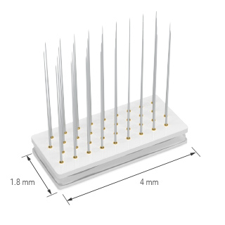 |
FMA 18-channel Electrode Array |
|
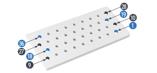
36 hole ceramic substrate with 400 µm hole separation |
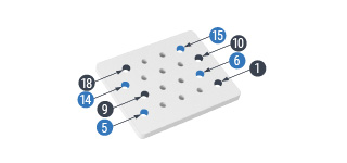
18 hole ceramic substrate with 400 µm hole separation |
|
Ceramic substrate zoom |
Ceramic substrate zoom |
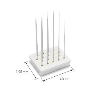
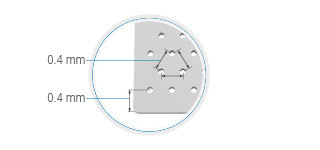
HD FMA
It features higher electrode density, reduced spacing between electrodes from 400 to 250 µm. It also has a smaller substrate size.
FMA 36-channel Electrode Array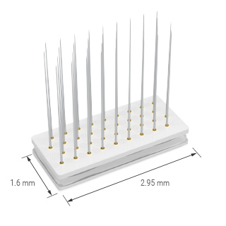 |
FMA 18-channel Electrode Array |
|
36 hole ceramic substrate with 400 µm hole separation |
18 hole ceramic substrate with 400 µm hole separation |
|
Ceramic substrate zoom |
Ceramic substrate zoom |
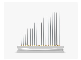
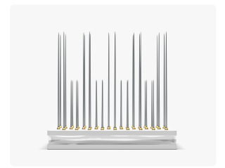
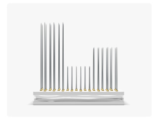
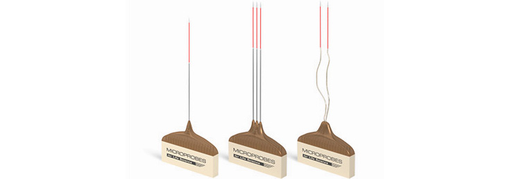
FMA Length Options
FMA are available in different length designs:
Uniform option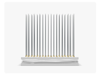 |
Slanted option |
|
Staggered option |
Clustered option |
FMA Technology
For over 50 years, Neuroscientists have routinely used metal microelectrodes inserted into the cortex and spinal cord to record and electrically stimulate neural elements. During this time, many electrode designs, ranging from single or bundled micro-wires to sophisticated silicon probes, have seen various successes in acute and chronic applications. For acute experiments, many neuroscientists typically fabricate bundles of micro-wires and insert them into the cortex using micro-drives.
As neuroscience research evolves to the study of large populations of cells in chronic rather than acute experiments, more sophisticated technologies must be employed to provide multi-electrode systems that can satisfy a diverse scope of experimental paradigms. Chronic experiments, which are conducted over months or even years, will require the use of intracortical microelectrodes for reliable neural interfaces for both stimulation and recording paradigms.
The need for arrays to have flexible design characteristics will be necessary to accommodate the varied experimental paradigms and animal models used among neuroscience researchers. Multi-electrode arrays that may have regular or irregular electrode-to-electrode geometric spacing, with multiple electrode depths that can stimulate or record from neurons without causing tissue damage or deterioration of the electrodes, are becoming essential tools. Even in the peripheral nervous system, emerging studies are investigating arrays of electrodes inserted into the spinal cord or nerve branches, with irregular electrode spacing, depth, and metal type as a means of providing a more sophisticated artificial neural interface.
Current research on neural prosthesis applications, including cochlear nucleus stimulation for an auditory prosthesis, cortical stimulation for a visual prosthesis, and cortical recording for brain-machine interfaces, all require use of electrode arrays maintained in a stable mechanical position relative to the associated neuronal structures.
MicroProbes has commercialized this innovated Floating Microelectrode Array (FMA) whose design permits the mixing of electrode types, impedance values, irregular electrode spacing, arbitrary electrode lengths, and electrode metals such as Platinum-Iridium and activated-iridium-oxide, within the same microelectrode array.
There are many investigative paradigms that require electrodes contained within a single array to have a range of tips exposures, as often characterized by their impedance, or a variety of electrode shaft lengths. Sometimes recordings are performed in a differential mode, requiring a reference electrode, which typically has an impedance value that is required to be an order of magnitude less than the recording electrodes.
“Ground” or “common” electrodes are also required to be included in both recording and stimulation multi-electrode arrays. Also, it is often desirable to implant an array along a sulcus, where some of the electrodes need to be much longer along the sulcus and shorter away from the sulcus.
MicroProbes' arrays are fabricated from biocompatible materials: alumina ceramic, Parylene-C, noble metals (gold, and platinum/iridium 70 % / 30 % or pure iridium), and medical implant grade silicone elastomers. Their FMAs implanted into animals have exhibited single unit activities for periods of over to 3.5 years. Moreover, researchers could record single unit activity from the same neuron for 1 month because of the floating nature of our FMAs.
MicroProbes' FMA design incorporates solid core conductors instead of silicone technology for several reasons. First, as a result of our initial research with the Visual Prosthesis Program at the Illinois Institute of Technology, (directed by Dr. Phil Troyk), an electrode design was required that could withstand indefinite stimulation without compromising the metal conductor or the integrity of the insulation interface. To date, metalized silicone probes have not demonstrated sufficiently robust behavior to warrant long-term stimulation. Secondly, their work with Dr. Richard Andersen’s laboratory at Cal Tech required floating microelectrode array designs that would accommodate electrode lengths up to 8 mm.
Researchers there also expressed the desire to have electrodes with different lengths within the same array. Microprobes has worked with these groups and others to develop a very flexible array design that is also very affordable for most laboratories.
Publications:
End-Stopping Predicts Curvature Tuning along the Ventral Stream
Neurons in primate inferotemporal cortex (IT) are clustered into patches of shared image...
Grasp Movement Decoding from Premotor and Parietal Cortex
Despite recent advances in harnessing cortical motor-related activity to control computer cursors...
In-Vivo Tests of a 16-Channel Implantable Wireless Neural Stimulator
Wireless stimulation of neural tissue could enable many emerging neural prosthesis designs, and...
Methodological considerations for a chronic neural interface with the cuneate nucleus of macaques
While the response properties of neurons in the somatosensory nerves and anterior parietal cortex...
Neuronal Prediction of Opponent’s Behavior during Cooperative Social Interchange in Primates
A cornerstone of successful social interchange is the ability to anticipate each other’s...
How to order:

Please contact the manufacturer, MicroProbes directly for further specifications and ordering. We would appreciate if you mention "Science Products" at any order or inquiry.
Online design forms
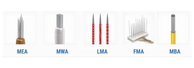
Related Products:
|
Floating Microelectrode Arrays (MicroProbes) |
(MicroProbes) |
(MicroProbes) |
|
(MicroProbes) |
Pt/Ir (MicroProbes) |
Pt/Ir (MicroProbes) |
|
Optogenetic Pt/Ir (MicroProbes) |
(PDF) |


-
Startseite
-
Produkte
-
Datenerfassung und -Analyse
-
Multielektroden-Aufnahmesysteme
-
Multikanal-Elektrodenträger für in-vivo-Ableitungen
- Floating Microelectrode Arrays (FMA)