Multi-Wavelength Beam Combiners for Mightex LED Collimator Sources
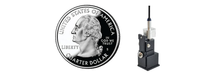
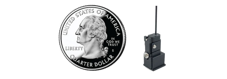
Mightex Multi-Wavelength beam combiners combine two LED collimators of different wavelengths into a single collimated beam. Multiple combiners can be cascaded to combine more than two LED collimator sources. At the heart of the beam combiner is a high-performance dichroic beam splitter that combines two wavelengths with > 95 % efficiency. A neutral beam splitter is also available as a lower-cost solution for applications where maximum light throughput is not required.
Features
- Combines two LED collimator sources into a single collimated beam
- Cascadable for more than two sources
- Precision locking tilt adjustment on each port
- High-efficiency dichroic beam splitters
- Low-cost neutral beam splitters available
- Integrated filter well for each beam
- Wide range of available wavelengths
- Multiple mounting features for lab and OEM applications
- Microscope adapters available
Applications
- Illumination for photonics applications
- Fiber coupling w/ optional focusing module
- Microscope illuminator
- General purpose beam combiner or splitter
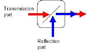
Beam combiners are characterized by the edge wavelengths of their dichroic beam splitters. Please use the following diagram and check the Specification table to select the correct beam combiner for intended LED collimator source.
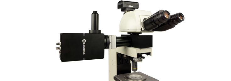
The beam combiner includes multiple mounting holes and threads so that it can be easily integrated into a system. Each input port features a fine 2-axis tilt adjustment to allow precise alignment of the LED collimator sources relative to system optical axis. A robust locking mechanism is also provided to maintain alignment over time. Additionally, adapters are also available for coupling to major brands of microscopes.
Adding optical filters to the input ports
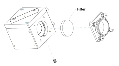
A filter well (see diagram below) is integrated into each input port so that a φ 1” or φ 25 mm optical filter can be inserted in between the LED source and the beam splitter. Narrow band filters and polarizers may be used to clean up LED spectrum or change the polarization state of the output beam. Here are the dimension requirement for the dichroic/beamsplitters to be fit into the beam combiner cubes:
- Length: 34~36 mm
- Width: 25~26.5 mm
- Thickness: 1mm recommended, 1.5 mm max.
Connecting multiple beam combiners
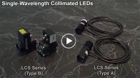
Multiple beam combiners can be connected using connecting plates (sold separately). See below video for example. A full instruction on how to build a Multi-Wavelength Collimated LED Sources can also be found at the manufacturer's website.
Connecting 11-mm aperture collimated LED sources
Currently, Mightex’s multi-wavelength beam combiners can only be used to DIRECTLY combine 22 mm-diameter Collimated LED Sources. For 11 mm-diameter collimated LED sources, (as shown below) a mechanical adapter (P/N: ACC-BC25-011) can be used to attach the LED sources to the beam combiners, before the 11-mm diameter collimated LED sources can be combined. We don’t yet have beam combiners for 38 mm- and 48 mm-diameter collimated LED sources.
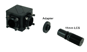
parts
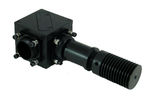
Specifications
Download Datasheet from manufacturer's website.
About the BLS-series BioLED light sources
Mightex BLS-series BioLED light sources are modularized fully-customizable turn-key solutions for optogenetics, fluorescence excitation, and other biophotonics applications. Precisely-timed and high-intensity light pulses are required in optogenetics experiments to activate channelrhodopsins (ChR2, ChR1 etc.) and halorhodopsins (NpHR) in order to excite and inhibit neurons. To meet these requirements, Mightex has developed a proprietary “IntelliPulsing” technology to allow BLS-series sources to output significantly higher power in pulse mode than what the LEDs are rated for in CW mode.
Features:
- High-power UV/VIS/NIR/white fiber-coupled LED’s
- Interchangeable fiber with SMA connector
- No moving parts in optical path
- Multiple mounting features for lab and OEM applications
- Optional LED controllers
- Compact, machined metal housing with integrated heat sink
- Locking electrical connector
Specification Sheet (in manufacturer's website)
Selecting a Microscopy LED Light Source
1. Select LED Wavelength
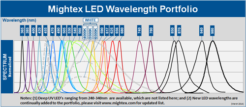
2. Select LED Configuration
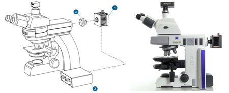
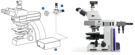
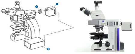
3. Select LED Controller
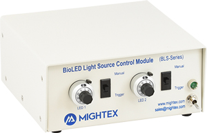
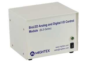
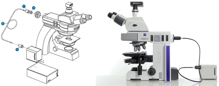
4. Microscope Coupling
Microscope adapters are available for all major manufacturers (Nikon, Olympus, Leica, Zeiss). If you have a different microscope, please contact us.

LED Application Examples
Abdelfattah AS et al., Bright and Photostable Chemigenetic Indicators for Extended In Vivo Voltage Imaging – Science (2019)
Abdelfattah et al., developed novel chemigenetic indicators for in vivo voltage imaging in neurons. To illuminate their indicators, the group used Mightex’s Microscope LEDs.
Crandall SR, et al., Infrabarrels Are Layer 6 Circuit Modules in the Barrel Cortex that Link Long-Range Inputs and Outputs – Cell Reports (2017)
In this work, Crandall et al., used Mightex’s Microscope LEDs to optically stimulating different neuronal pathways connecting to pyramidal neurons found in L6 of the whisker somatosensory cortex of mice.
Higgs MH, Measurement of Phase Resetting Curves Using Optogenetic Barrage Stimuli – Journal of Neuroscience Methods (2017)
To study phase resetting curves in vivo, Higgs and Wilson used Mightex’s Microscope LEDs to establish whether their novel method of optogenetic barrage stimulation would work during extracellular spike recordings (a recording method often used in in vivo experiments).
Also See:
Related Products:

